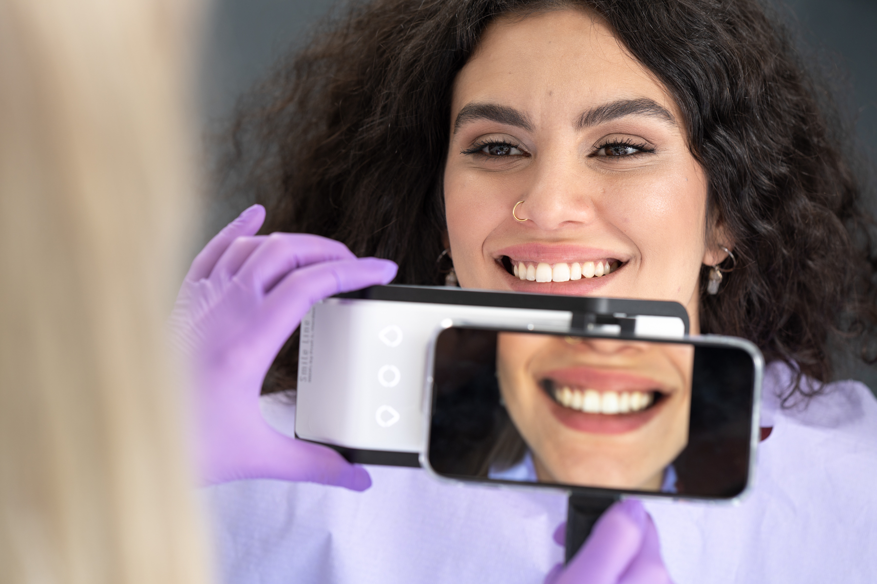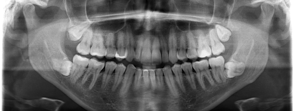
An orthopantomogram is a type of dental x-ray used to get a full overview of the teeth and surrounding bone structures. It is commonly done at the first dental visit and is also recommended during annual check-ups.
It is especially useful when a complete view of all teeth is more important than detailed images of individual ones.
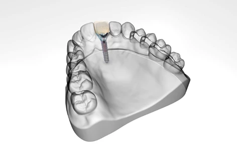
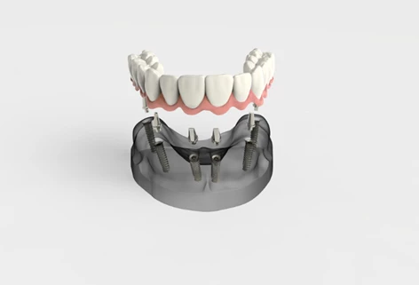
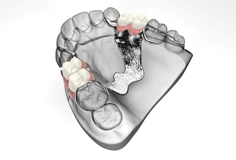
In addition to being an orientation scan, an orthopantomogram is used in planning prosthetic treatments.
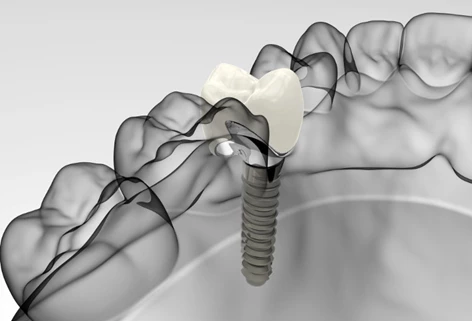
It is also used in orthodontics and oral surgery - for implant planning or assessing unerupted teeth.
Digital panoramic x-rays are the most common type. They involve minimal radiation exposure and offer significantly improved accuracy and diagnostic value compared to conventional imaging.
Thanks to modern digital technology, our panoramic dental x-ray uses the lowest possible radiation dose, which is considered non-harmful.
The imaging is safe for children - the dosage is adapted to be even lower.
We use the advanced Carestream CS 8200 digital system, which ensures minimal exposure.
The only group advised to postpone x-rays are pregnant women, especially during the first trimester. In such cases, we recommend waiting until after delivery.
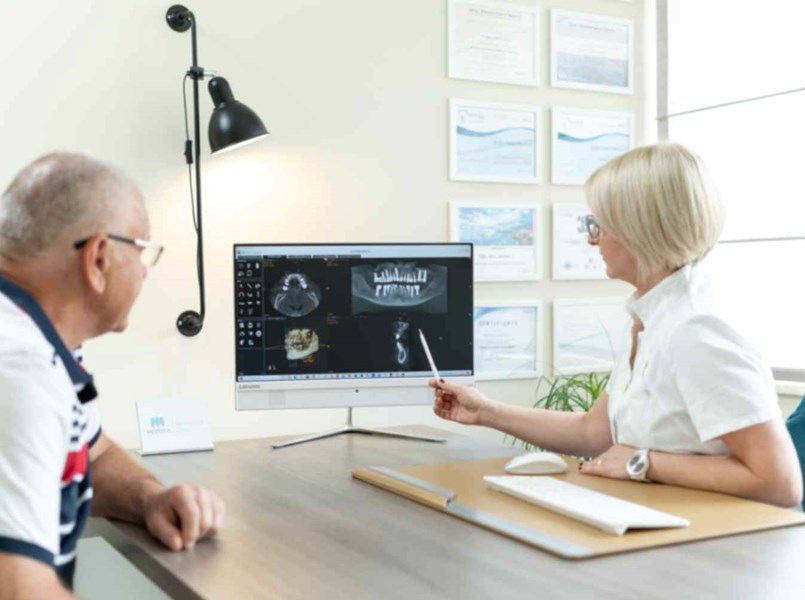
An orthopantomogram is crucial for establishing a correct diagnosis and planning treatment steps.
Our advanced system provides top-quality dental x-rays, enabling the most accurate diagnosis possible.
Before entering the imaging room, the patient must remove all metal objects such as earrings, necklaces, nose or tongue piercings, and any other jewelry, as well as take out any removable dental prostheses (dentures).
Metal objects like jewelry and dentures must be removed because they can cause artifacts on the x-ray image, which may interfere with or prevent accurate diagnosis.
After that, a lead apron is placed on the patient to protect against radiation, and they are properly positioned in front of the panoramic x-ray machine to ensure the best possible image quality.
During the scan, the patient is in a specially shielded room with lead-lined walls, while the device moves around their head to capture a full view of the teeth, jaws, and surrounding structures.
It is important to remain still and avoid any movement during the panoramic x-ray.
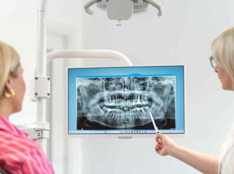
Immediately after the scan is completed, the orthopantomogram is processed and delivered to the patient via Viber or email.
This ensures quick and easy access to the data needed for further dental analysis and treatment.
If you happen to lose the x-ray, you can always contact us to have it resent.
The price of a panoramic x-ray (orthopantomogram) usually ranges from €25 to €50. When choosing where to get a dental x-ray, many patients focus solely on the price, forgetting how important the image quality is.
While cost is a relevant factor, the precision of the x-ray device plays a key role in ensuring an accurate diagnosis and successful treatment.
At our clinic, we use the Carestream CS 8200, a high-end orthopantomogram unit with a resolution of 75 microns - among the most precise on the market. It produces detailed images that can mean the difference between saving or losing a tooth.
For example, a less accurate scan might suggest a root infection and lead to unnecessary tooth extraction, while our detailed image may reveal it’s just a thinning of the bone - meaning extraction is not needed.
Price differences in x-rays mostly result from the quality of equipment used. High-resolution devices cost up to twice as much as standard ones, but offer significantly better diagnostic reliability.
Choosing a cheaper x-ray without considering the accuracy of the scan can lead to misdiagnosis and even tooth loss. Saving €10 or €15 is not worth the risk if it affects treatment decisions.
We recommend having a panoramic x-ray once a year. Daily stress and a fast-paced lifestyle can negatively affect your oral health without any visible symptoms.
Significant changes in bone or teeth can occur in just 12 months - and an annual dental x-ray can help detect problems early and prevent complications.
The main benefit of a panoramic x-ray is early detection. It can reveal cavities, cysts, infections, root changes, or bone loss before any symptoms appear. This makes treatment planning easier and more accurate.
If your dentist notices anything unclear on the orthopantomogram, they might recommend a 3D scan of the jaw (CBCT), which gives even more detailed insight into tooth and bone structures.
CBCT is used in complex cases, such as implant planning, unclear diagnoses, or to evaluate anatomical structures before surgery.
Although both panoramic x-rays (orthopantomograms) and CBCT scans are used to examine the jaw, they serve different purposes and offer different types of information.
A panoramic x-ray is a two-dimensional image that gives a general overview of the teeth, jawbone, joints, and surrounding tissue. It is quick, involves low radiation, and is usually the first step in diagnosis. It helps detect decay, bone changes, root issues, and abnormal tooth positions.
CBCT, on the other hand, creates a three-dimensional view of the same structures. It is used when more detail is needed, especially for implant planning, root canal treatment, detecting hidden canals, or checking the relationship between teeth, nerves, and sinuses. It uses more radiation than a panoramic x-ray but much less than medical CT.
In short - panoramic x-rays give us a wide overview, while CBCT allows us to look at structures in detail, layer by layer. A good dentist will know when which method is appropriate.
A referral is not always necessary for a panoramic dental x-ray, but there must be a justified medical reason. Since this is a radiological procedure, it should be based on clinical judgement.
If you do not already have a recommendation for an orthopantomogram, our dental team can examine you on-site and advise whether it is needed. If so, they will let you know when the scan should be done.
A panoramic x-ray shows the condition of your teeth, jawbone, and surrounding structures. It is often requested as part of preparation for orthodontic treatment, implant planning, endodontic therapy, or in case of pain, swelling, or suspected infection.
In the end, a panoramic x-ray is not an administrative step, but a clinical decision - and should be taken only when truly necessary.