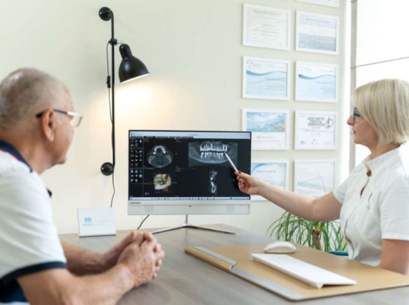
Diagnostics and oral health monitoring play a key role in maintaining overall health. In this context, a CT (computed tomography) scan of the jaw becomes an irreplaceable tool in identifying and tracking various diseases and conditions of the oral cavity.

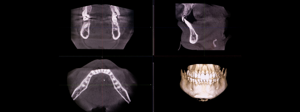
CBCT scan of the teeth and jaw uses x-rays to produce a detailed three-dimensional image of oral structures. This technology allows accurate visualisation of bones, teeth, sinuses, nerves, soft tissue, and other anatomical features.
The result is a detailed image that aids in problem identification and treatment planning.
It has become an invaluable tool in dentistry and oral surgery. Its ability to detect and clearly visualise various oral conditions enables earlier diagnosis and more effective therapy.
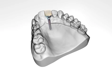
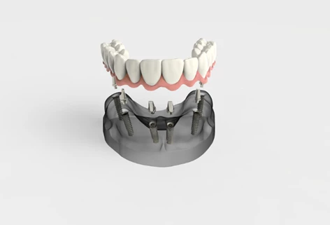
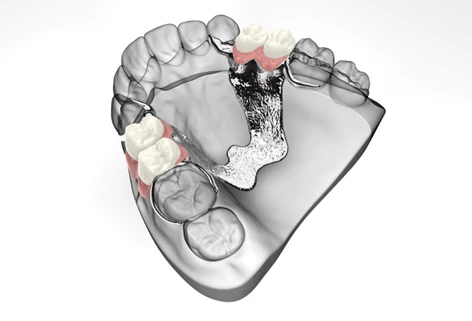
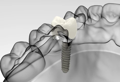
For dental implants placement, we can determine the optimal position of each implant. We need to be sure the implant will not interfere with vital anatomical structures such as the inferior alveolar nerve, sublingual artery, maxillary sinus, or nasal cavity. This approach helps prevent serious surgical complications. Possible risks include temporary or permanent numbness in the lips and potentially severe bleeding.
The CBCT scanner is adjusted depending on the area that needs to be scanned.
The patient gently bites into a special holder to ensure correct jaw positioning.
The device rotates around the patient’s head, typically for 10 to 20 seconds, capturing multiple images from different angles.
It is important the patient remains still and avoids swallowing to ensure a clear and sharp scan.
Orthopantomogram (panoramic dental x-ray) and CBCT (cone beam computed tomography) are two different radiographic techniques used to image the teeth and jaw, each for different purposes.
An orthopantomogram provides a two-dimensional (2D) view of the entire upper and lower jaw, including teeth, bones, and surrounding structures. It’s used for basic diagnostics, assessing dental health, detecting cavities, bone condition, and the presence of cysts or impacted teeth.
On the other hand, a CBCT scan provides a three-dimensional (3D) image of the jaw and teeth with a high level of accuracy.
Thanks to its detailed imaging, it’s used in more complex cases such as planning implant procedures, root canal therapy, and diagnosing temporomandibular joint disorders.
Unlike a panoramic x-ray, the dental cone beam CT creates layered images that give the dentist better spatial orientation and more precise diagnostics.
The main difference is that while a panoramic x-ray offers an overview in 2D, the CBCT scan delivers a detailed 3D view of the jaw and teeth, which is essential for planning more advanced dental treatments.
The price of a CBCT scan at Centrodent depends on the scope of imaging:
Our Carestream CS 8200 3D CT scanner operates at an extremely high resolution of 75 microns, which is among the highest available in dental diagnostics.
This high image quality allows for a detailed view of fine anatomical structures, which is essential for accurate diagnostics and therapy planning.
In addition, the device uses MAR technology (Metal Artifact Reduction), which minimizes distortions and noise caused by metal presence, such as implants, amalgam fillings, or metal restorations.
This means clearer and more precise images, giving our dentists a better view of the bone and teeth condition without visual interference.
CT scan stands for Computed Tomography scan.
It is a medical imaging technique that uses computer-processed combinations of multiple X-ray measurements taken from different angles to produce cross-sectional (tomographic) images of specific areas of the body. This allows doctors to see inside the body in much more detail than standard X-rays.